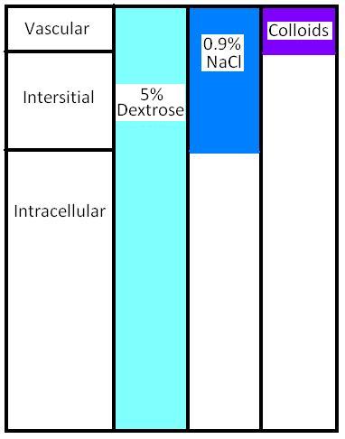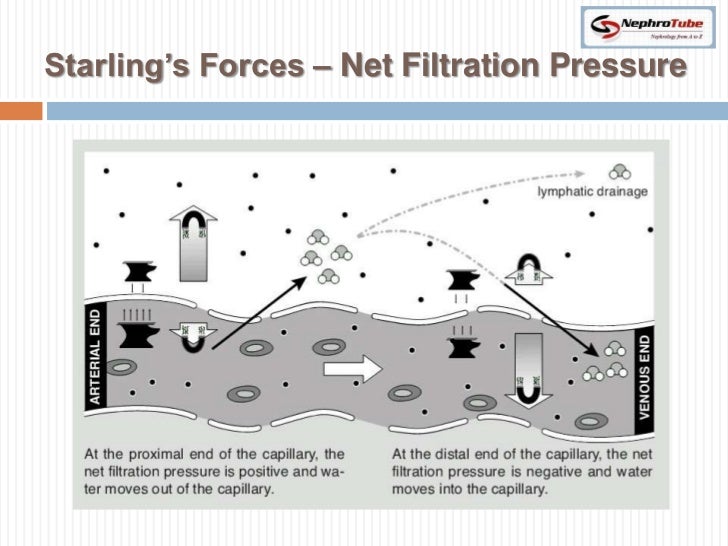
These figure are not to scale and thus the reader should not try to interpret the quantitative values of the osmolarity and volume, but should focus on their qualitative increase or decrease. In the case of secretory diarrhea, no change in ECF osmolarity is caused and thus there is no shift in fluid between the ICF and ECF.

As seen, the driving force for the shifting fluid is a change in the ECF's osmolarity. The initial change from normal is given above followed by the compensatory body fluid shift below. the volume (x-axis) of the body's ICF and ECF during states of pathological volume loss. These graphs provide a representation of the osmolarity (y-axis) vs. Once the ECF osmolarity changes, water shifts into or out of the intracellular compartment, within minutes, until a new osmolar equilibrium is achieved between the ECF and ICF. Solutes and water can be added to it from the GI tract whereas they can be subtracted from it by a variety of processes, most notably by the action of the kidneys but also by loss from perspiration, breathing, and the feces.


 0 kommentar(er)
